
Dental X-rays, or radiographs, have long been an essential diagnostic tool in the world of dentistry. Over the years, advances in X-ray technology have transformed the way dental professionals diagnose and treat oral health issues. While the primary function of X-rays was initially cavity detection, the scope of their application has grown to encompass a wide range of issues, including bone health, jaw alignment, tooth development, and even the identification of tumors or cysts.
In this in-depth article, we will explore how modern dental X-rays work, their different types, their role in detecting cavities, assessing jaw health, and their essential function in a variety of dental specialties, from general dentistry to orthodontics. We will also delve into the benefits of modern dental X-rays and how they contribute to better treatment outcomes and patient safety.
How Modern Dental X-Rays Work: The Science Behind the Digital Images
Dental X-rays are an indispensable tool in the dentist’s diagnostic toolkit. X-rays use a controlled beam of radiation to capture images of the inside of the mouth, revealing the structure of the teeth, gums, and bone. The procedure works by directing the X-ray beam through the mouth, while a film or digital sensor on the other side records the image.
The key principle of dental X-rays is the difference in how various tissues absorb radiation. Dense tissues like teeth and bones absorb more radiation, appearing white or light gray on the X-ray image, while softer tissues like gums and muscles allow more radiation to pass through, appearing darker on the image.
The captured X-ray image is then either processed on film or digitally, depending on the type of system being used. Film-based X-rays require chemical processing, whereas digital X-rays are displayed almost instantly on a computer screen. The images generated from X-rays provide valuable insights into the patient’s oral health, helping dentists identify issues that may not be visible to the naked eye.
The Different Types of Dental X-Rays
Dental X-rays come in several forms, each serving a distinct purpose in diagnosing oral health problems. The most common types include bitewing, periapical, panoramic, and cone beam computed tomography (CBCT) scans. Each type of X-ray offers unique benefits depending on the issue being evaluated.
1. Bitewing X-Rays: Detecting Cavities Between Teeth
Bitewing X-rays are the most commonly used type of dental X-ray. These images are taken by having the patient bite down on a small piece of film or a digital sensor. Bitewing X-rays provide a clear view of the crowns of the upper and lower teeth, especially those in the back of the mouth.
Bitewing X-rays are most effective at detecting cavities between the teeth, which are often difficult to see during a regular dental exam. The X-ray captures the spaces between the teeth, revealing early-stage tooth decay before it becomes visible on the surface. Early detection allows for prompt intervention, such as fillings or preventive care, which can prevent further damage and avoid more invasive procedures down the road.
Additionally, bitewing X-rays allow dentists to assess bone levels in the areas surrounding the teeth. This is crucial for detecting early signs of gum disease, as bone loss is a key indicator of periodontal issues.
2. Periapical X-Rays: Comprehensive Tooth and Root Assessment
Periapical X-rays capture the full view of an individual tooth, from the crown to the root, as well as the surrounding bone. These X-rays are typically taken when a dentist suspects an issue with the root or bone beneath the tooth’s surface. Periapical X-rays are particularly useful for detecting abscesses, cysts, or infections that are affecting the tooth root.
A periapical X-ray is invaluable for assessing the health of the tooth’s root, the supporting bone structure, and the surrounding tissues. These X-rays help identify problems such as tooth abscesses, which can lead to infection and potential tooth loss if not treated promptly. They are also used to detect deep cavities, root fractures, and other abnormalities that may not be visible through visual examination alone.
3. Panoramic X-Rays: A Full-Mouth Overview
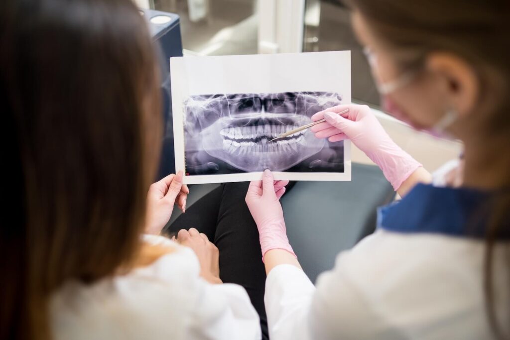
Panoramic X-rays provide a comprehensive, broad view of the entire mouth, including the upper and lower jaws, teeth, sinuses, and surrounding structures such as the temporomandibular joints (TMJ). This type of X-ray is useful for detecting large-scale issues like impacted teeth, jawbone irregularities, tumors, or cysts.
The panoramic X-ray is especially helpful in planning for more complex dental treatments, such as implants, extractions, or orthodontic procedures. It allows the dentist to assess the overall structure of the mouth and determine how best to proceed with treatment. Panoramic X-rays are also frequently used to evaluate the position of wisdom teeth, which are often a source of dental complications.
4. Cone Beam CT (CBCT) Scans: 3D Imaging for Complex Cases
Cone beam computed tomography (CBCT) is an advanced imaging technique that produces high-quality 3D images of the teeth, jawbone, and soft tissues. Unlike traditional X-rays, which offer two-dimensional images, CBCT provides a more detailed view of the oral structures, making it ideal for complex diagnostic cases.
CBCT is commonly used in implant dentistry, where precision is critical for determining the best placement of dental implants. It is also used in evaluating TMJ disorders, assessing bone structure for orthodontic treatments, and planning for surgeries that involve the jaw or teeth. The ability to see the teeth and surrounding tissues in three dimensions allows for better planning and more accurate outcomes.
Why Dental X-Rays Are Vital for Detecting Cavities and Other Dental Issues
Cavities, or dental caries, are one of the most common dental problems faced by individuals worldwide. While cavities often begin as small, localized areas of enamel erosion, they can quickly progress into larger, more serious problems if not detected and treated early. Modern dental X-rays play a crucial role in identifying cavities, especially those that are hidden between teeth or beneath existing dental work.
Early detection of cavities is essential for preventing the need for more invasive treatments, such as root canals or extractions. With the help of bitewing X-rays, dentists can identify early stages of decay before the damage is visible on the surface. By catching cavities early, dental professionals can intervene with a simple filling rather than a more costly and painful procedure down the road.
Dental X-rays also allow dentists to assess the severity of existing cavities and determine whether they have spread to the tooth’s pulp or root. This is particularly important in determining whether a root canal or other restorative procedure is necessary.
Assessing Jaw Health and Bone Density
The jawbone is the foundation that supports our teeth, and its health is essential for maintaining a functional bite and overall oral health. Periodontal disease, tooth loss, and trauma can lead to the gradual breakdown of bone density, making the teeth less stable and increasing the risk of further damage.
Dental X-rays are invaluable tools for evaluating the health of the jawbone and surrounding structures. Periapical and bitewing X-rays allow dentists to assess bone levels and identify early signs of bone loss. This is especially important for detecting periodontal disease, a condition in which the gums become infected and the bone supporting the teeth begins to deteriorate.
For patients with missing teeth, dental X-rays help determine the need for bone grafts or dental implants. If the bone density is insufficient, the dentist may recommend procedures to restore bone mass before placing implants.
The Role of Dental X-Rays in Diagnosing TMJ Disorders
The temporomandibular joint (TMJ) is the hinge that connects the jaw to the skull, and disorders of the TMJ can cause pain, clicking, or difficulty moving the jaw. TMJ disorders can be caused by factors such as teeth grinding, arthritis, jaw misalignment, or trauma. While a dentist or orthodontist can often diagnose TMJ issues based on symptoms and physical examination, X-rays provide a more detailed view of the joint and surrounding structures.
Panoramic X-rays are frequently used to assess the alignment and condition of the TMJ, helping to identify issues such as joint inflammation, bone erosion, or displacement. In more complex cases, a CBCT scan can provide a detailed 3D image of the jaw and TMJ, helping the dentist or specialist plan appropriate treatment, which may include physical therapy, splints, or even surgery.
The Safety of Modern Dental X-Rays
One of the most common concerns patients have when it comes to dental X-rays is the potential for radiation exposure. However, modern dental X-ray technology has made significant strides in minimizing radiation exposure, especially with the advent of digital X-rays. Digital X-rays emit up to 80% less radiation than traditional film-based X-rays, making them much safer for patients.
Additionally, dental professionals take precautions to ensure that patients are protected from unnecessary radiation. Lead aprons and thyroid collars are commonly used during X-ray procedures to shield vital organs from radiation. The amount of radiation used in dental X-rays is very low, and for most patients, the benefits of X-ray imaging far outweigh the minimal risks.
Conclusion: The Unmatched Power of Dental X-Rays in Oral Healthcare
Modern dental X-rays have transformed the way dentists diagnose and treat dental conditions, providing unparalleled insights into the health of the teeth, gums, and jaw. Whether it’s detecting cavities in their earliest stages, assessing bone health, evaluating TMJ disorders, or planning for orthodontic or implant procedures, dental X-rays are an indispensable tool in ensuring optimal oral health.
Advancements in digital X-ray technology have made imaging faster, more accurate, and safer for patients. With lower radiation exposure, quicker image processing, and clearer visuals, digital X-rays are the gold standard in modern dental care. By embracing the power of X-rays, dental professionals can offer more precise diagnoses and personalized treatment plans, ultimately leading to healthier smiles and more satisfied patients.
For those looking to maintain or restore their oral health, visiting a dental clinic equipped with the latest X-ray technology is an investment in both your peace of mind and the long-term health of your teeth and jaw. Whether it’s for routine exams, cavity detection, or more complex treatments like implants or TMJ therapy, dental X-rays are an invaluable resource for comprehensive care.
As dental technology continues to evolve, the future of dental X-rays promises even more exciting developments, such as enhanced imaging capabilities and even lower radiation doses.
For now, dental X-rays remain an essential component of comprehensive oral care, helping patients maintain the health of their teeth, gums, and jaws for years to come. To experience the benefits of modern dental diagnostics, consider scheduling an appointment with a trusted dental provider who utilizes the latest X-ray technologies to ensure your oral health is in good hands.